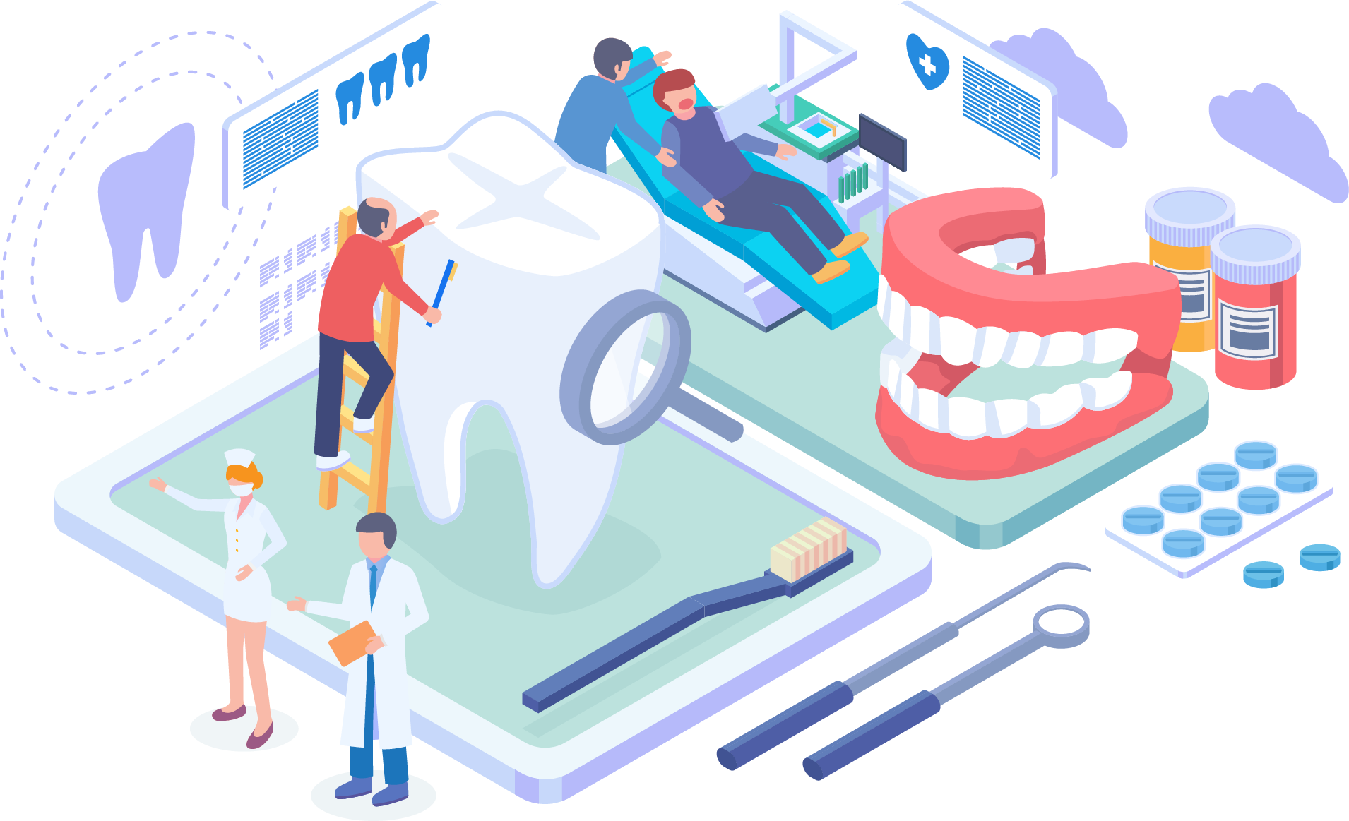
Dental X-Rays
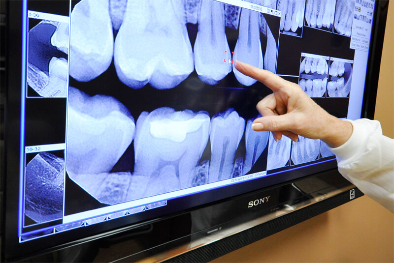
Dental imaging refers to the use of x-ray images (radiographs) to visualise the dentition and its surrounding structures. Radiographic technology has changed significantly over the years and most modern dental practices would now be equipped with digital units which require lower effective doses.
Higher end equipment are often capable of producing both 2D as well as 3D images. Dental radiographs are sometimes necessary for the diagnosis and detailed examination of teeth and jaws prior to treatment. Our digital radiography system provides a lower dosed x-ray for patients and is more time efficient in developing and processing of x-rays.
Dental X-Rays
At NoFrills Dental, we offer a variety of dental x-rays for our patients. With these tools at our disposal, our clinical teams are fully equipped to serve your dental needs. There are many kinds of dental x-rays that we use; periapical, bitewing, occlusal, cephalogram, panoramic, cone beam computer tomography.
PERIAPICAL VIEW
BITEWING VIEW
OCCLUSAL VIEW
FULL MOUTH SERIES
CEPHALOGRAM
PANORAMIC (ORTHOPANTOMOGRAM)
CONE BEAM COMPUTED TOMOGRAPHY (CBCT SCAN)
Benefits of Digital X-Rays
With the use of x-rays, the dentist will be able to locate hidden dental structures (e.g. roots of teeth, buried teeth), establish bone levels and detect disease (e.g. cavities, benign or malignant masses, bone loss) that are otherwise not visible to the dentist.
Treatment Process
The x-ray view is dependent on the location, size and structure of the ‘area of interest’ to the dentist. Smaller images often require for the film/sensor to be placed in the mouth (collectively known as intra-oral views) while larger images require the film/sensor to be located outside the mouth (collectively known as extra-oral views).>
Are Dental X-rays Safe?
An X-ray image is formed when a controlled release of X-ray radiation penetrates the oral structures before striking a film or sensor. Dense objects (e.g. teeth and bone) impedes x-ray radiation penetration appear lighter as less radiation will strike the film or sensor. Dental caries and soft tissues appear darker as they are less dense and are easily penetrated by the X-rays. Dental restorations (fillings, crowns) may appear lighter or darker, depending on the density of the material used in their fabrication.
The X-ray radiation dosage received by a dental patient is typically very small, equivalent to a few days’ worth of background environmental radiation, or an airplane flight between Singapore and Shanghai. The beam is concentrated into one short burst aimed at a small area and incidental exposure of other parts of the body is further reduced by the use of a lead apron.
Traditionally, exposed x-ray films is developed using a series of chemicals in a dark room. The adoption of electronic x-ray radiation sensors have made this process faster, more environmental friendly and reduce the effective radiation dosage, making it safer for patients and staff.
Ready to redesign your smile?
Let us help you through your dental journey.